Describe the Layers of the Heart Wall
It contracts to pump blood out of the heart and then relaxes as the heart refills with returning blood. The inner layer of the heart.
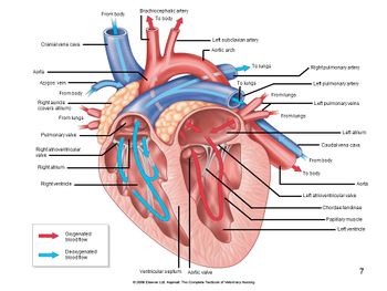
The Heart Circulatory Anatomy
The chambers of the heart.
. The wall of the heart is composed of three layers of unequal thickness. It is the outermost layer. It is the middle layer.
It lines the cavities and valves of the heart. The layers of the heart are as follows. Describe the three layers of the heart wall.
Answer 1 Coverings and layers of the heart wall Pericardium is the outer covering of the heart. Describe the layers of the pericardial sac. The outer layer of the wall of the heart.
It is the thickest and made up of cardiac muscles which are striated but involuntary. Describe the three layers of the heart. The muscles of thethigh are composed of skeletal muscle tissue.
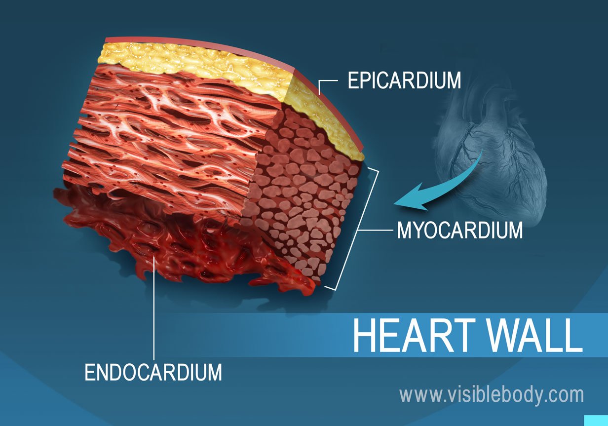
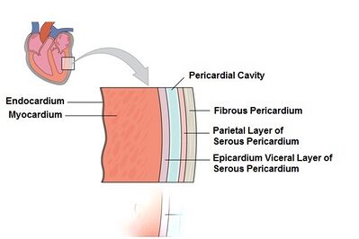
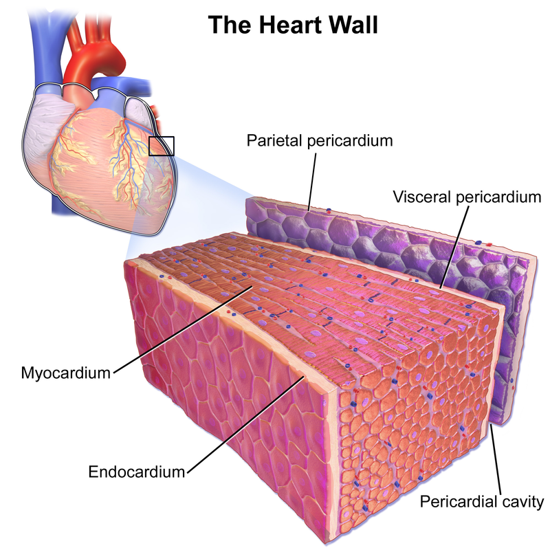
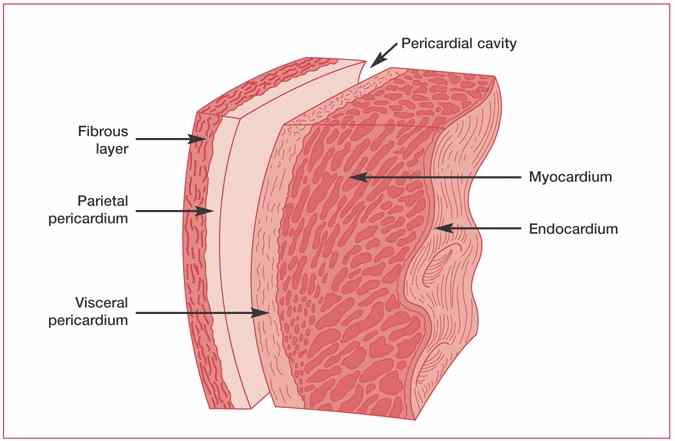
From superficial to deep these are the epicardium the myocardium and the endocardium. Endocardium- Covers inner surfaces of heart including valves. Myocardium- Middle muscular layer forming atria and ventricles.
The myocardium is the muscular wall of the heart or the heart muscle. The muscular middle layer of the wall of the heart. The outermost covering of heart is fibrous pericardium that protects the heart and it also h View the full answer.
The wall of heart is made up of following three layers from outside to inside. The heart wall consists of three layers. Epicardium Protective outer layer made of connective tissue covered by a thin epithelium.
Visceral layer of serous pericardium. In this picture above weve sliced the heart. Three Layers of the Heart Wall.
The anatomy of the heart. Layers of the Heart Wall. The heart chamber is the space inside the heart and thats normally filled with blood.
It preforms the function of pumping what is necessary for the circulation of blood. The great vessels of the heart. This is the outermost layer of the heart and is one of the two layers of the pericardium.
Structure of the Heart. Outer layer of the heart and inner most layer of the serous pericardium. Composed of mesothelium and adipose tissue.
Epicardium visceral layer Describe the structure and function of each of the three layers of the heart wall. The outermost layer of the wall of the heart is also the innermost layer of the pericardium the epicardium or the visceral pericardium discussed earlier. This thin layer.
The esophageal wallincludes a middle layer of dense irregular connective tissue. Endocardium The innermost layer of the cardiac wall is known as the endocardium. The valves of the heart.
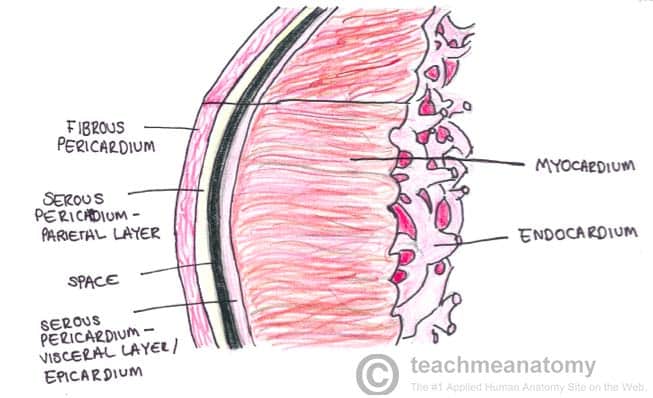
The Heart Wall - TeachMeAnatomy. Epicardium- Covers surface of the heart. It lines the cavities and valves of the heart.
It is made up of simple squamous epithelium. Epicardium -the outside of the myocardium is covered with a thin layer called the epicardium Visceral layer of serous pericardium. The innermost layer of the cardiac wall is known as the endocardium.
All this red stuff is heart muscle called myocardium and then theres a membrane surrounding it and we call it the pericardium. Myocardium Muscular middle layer made of cardiac muscle whose fibres contract spontaneously to produce the heartbeat. The layers of the heart wall.
There are two type of pericardium. Which of the three layers is most important in causing contractions of the heart. The heart wall consists of three layers.
There arefenestrations openings in the epithelial cells of capillary walls. Find step-by-step Anatomy and physiology solutions and your answer to the following textbook question. Know all this a.
It is the most massive part of the heart. This is the middle layer of the heart that contains the cardiac muscular tissue. The heart is enclosed in a pericardial sac that is lined with the parietal layers of a serous membraneThe visceral layer of the serous membrane forms the epicardium.
A serous membrane that forms the innermost layer of the pericardium and the outer surface of the heart. A Fibrous pericardium. 4 rows Key facts about the layers of the heart.
The heart wall The heart wall itself can be divided into three distinct layers. Explain the anatomy of the tricuspid and bicuspid or mitral valves and their chordae tendineae and papillary muscles. Layers of the Pericardium Heart Wall and Spiral Arrangement.
Pericardium or the layer surrounding the heart Epicardium the outside layer of the heart Myocardium the middle layer of the heart Endocardium the innermost layer of the heart. Epicardium visceral pericardium. The walls of bloodcapillaries are composed of a thin epithelium.
The human heart is a four-chambered muscular organ shaped and sized roughly like a mans closed fist with two-thirds of the mass to the left of midline.

Layers Of The Pericardium Heart Wall And Spiral Arrangement Physiology Cardiovascular System Anatomy And Physiology

What Are The 3 Layers Of The Heart What Function Does Each Layer Serve Quora

Heart Anatomy Anatomy And Physiology I
Layers Of The Heart Video Organ Systems Khan Academy
How Many Walls Does The Human Heart Have Socratic

Pericardium And Layers Of Heart Wall Diagram Quizlet

The Three Layers Of The Heart Wall Location Structure Function Video Lesson Transcript Study Com

Heart And Great Vessels Anterior View The Heart Is Enclosed In The Pericardial Sac The Innermost Layer O Heart Structure Cardiovascular System Nursing Exam

The Three Layers Of The Heart Wall Location Structure Function Video Lesson Transcript Study Com

Epicardium What Is It Functions And More Osmosis
The Heart Wall Teachmeanatomy
Anatomy Of The Human Heart Physiopedia
What Is The Name Of The Double Layered Membrane That Surrounds The Heart Socratic
What Are The 3 Layers Of The Heart What Function Does Each Layer Serve Quora
Where Is The Visceral Pericardium Located Socratic

Anatomy And Physiology Of The Heart Springer Publishing

Heart Anatomy Anatomy And Physiology I
Heart Structure Anatomy Physiology Wikivet English

Pericardium Structure Function


Comments
Post a Comment